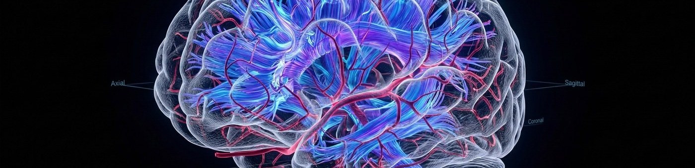
Neuroradiology Services
AI-Enhanced Brain, Spine & Head Reads
Neuroradiology Studies We Cover
Every study listed below is read by fellowship-trained neuroradiologists — not general radiologists with neuro experience, but dedicated subspecialists.
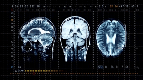
Brain MRI & CT
Stroke protocol, demyelinating disease, epilepsy, tumor, infection, and post-operative brain. Including DWI, ADC, perfusion, and spectroscopy sequences.
Spine MRI & CT
Cervical, thoracic, and lumbar spine — including herniation, stenosis, cord compression, myelopathy, infection, and post-surgical spine.
MR Angiography
Intracranial MRA, carotid MRA, circle of Willis, and cervicocerebral MRA. Aneurysm detection, stenosis assessment, and vascular malformation characterization.
CT Angiography (Neuro)
CTA head and neck for aneurysm, stenosis, and dissection. CT perfusion for stroke triage. Used in acute stroke workflow at many partner facilities.
Head & Neck Imaging
Temporal bone CT, paranasal sinus CT and MRI, orbit MRI, salivary gland, neck mass workup, skull base lesions, and inner ear anatomy.
Peripheral Nerve & Plexus
MR neurography, brachial and lumbosacral plexus MRI, nerve sheath tumor evaluation. High-resolution sequences reviewed by neuroradiologists trained in neurography.
Critical Protocols Built In
These clinical pathways are pre-configured at onboarding — no extra setup required.
Stroke Protocol
Acute stroke studies (non-contrast CT brain, CTA head and neck, CT perfusion) are auto-flagged STAT and routed to the next available neuroradiologist. Critical imaging findings are called within minutes of detection.
Intracranial Hemorrhage Alert
AI detection of intracranial hemorrhage on CT triggers immediate STAT escalation. The neuroradiologist is notified before they open the study. Your clinical team receives a phone call the moment the read is confirmed.
Mass Effect and Herniation
Studies showing mass effect, midline shift, or uncal herniation are escalated automatically regardless of original priority level. These are treated as critical findings under our standard workflow.
Neuroradiology Reading, by the Numbers
Key metrics across neuroradiology reads
< 2
min — AI hemorrhage detection on CT
< 45
min — STAT neuro study turnaround
6+
Neuro study types covered
100%
Fellowship-trained on every read
Subspecialty Neuroradiology Coverage
Frequently Asked Questions
Are your neuroradiology reads done by fellowship-trained radiologists?
Yes. All neuroradiology studies at Natoe AI are read exclusively by fellowship-trained neuroradiologists. We do not route neuro studies to general radiologists or to radiologists with self-reported neuro experience.
Can Natoe AI support our stroke program?
Yes. Our stroke protocol is pre-built: non-contrast CT brain, CTA head and neck, and CT perfusion studies are auto-flagged STAT and routed to the next available neuroradiologist. Critical findings are called immediately. Imaging centers that perform stroke protocol imaging can rely on Natoe AI for fast, fellowship-trained reads.
Do you read MR spectroscopy and advanced MRI sequences?
Yes. Our neuroradiologists read diffusion-weighted imaging (DWI), ADC mapping, MR perfusion (DSC and DCE), MR spectroscopy, susceptibility-weighted imaging (SWI), and functional MRI in clinical contexts.
Can Natoe AI cover head and neck imaging in addition to brain and spine?
Yes. Head and neck imaging — including temporal bone CT, paranasal sinus studies, orbit MRI, skull base, and neck mass evaluation — is covered under our neuroradiology service. These studies are read by neuroradiologists with head and neck subspecialty training.
What is turnaround time for neuroradiology studies?
STAT neuroradiology studies (stroke protocol, ICH, mass effect) are typically reported within 45 minutes. Routine neuro studies (outpatient brain or spine MRI) are reported within 4–6 hours. All reads are same-day, aligned with outpatient imaging center operating hours.
Are you hiring fellowship-trained neuroradiologists?
Yes. Natoe AI hires fellowship-trained, board-certified neuroradiologists for remote reading — subspecialty rates 25–40% higher than general radiology. See the Remote Neuroradiologist role for current details, or browse all open radiologist roles.