How Does Teleradiology Work? Step-by-Step Workflow for Imaging Centers (2026)
Reviewed by board-certified radiologists
Medically reviewed by: Natoe AI Clinical Team — Peer-reviewed internally — pending formal medical advisor appointment. Last reviewed: 2026-05-13.
Short answer: A teleradiology workflow has seven stages: (1) imaging study acquired at the imaging center, (2) study uploaded to the modality's PACS, (3) study transmitted via secure cloud or VPN to the teleradiology provider, (4) study routed by AI or routing rules to the right subspecialist radiologist, (5) radiologist opens the study on a diagnostic-grade workstation and dictates a structured report, (6) AI-assisted draft is reviewed and signed off by the radiologist, (7) signed report returns to the imaging center's RIS for the referring physician. Total turnaround typically ranges from 30 minutes for STAT studies to same-day for routine reads.
Why the Teleradiology Workflow Matters
For imaging center operators evaluating teleradiology providers, the actual workflow — not the marketing — determines turnaround time, report quality, and whether your reads slot cleanly into your downstream RIS or create friction. For radiologists evaluating teleradiology employers, the workflow determines how many studies per shift are realistic, the quality of the worklist routing, and how much AI tooling actually saves time versus adds cognitive overhead.
This guide walks through the full workflow stage-by-stage. For the conceptual definition and history of teleradiology, see our What Is Teleradiology guide.
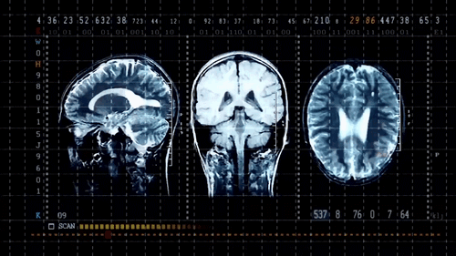
Stage 1 — Image Acquisition at the Imaging Center
The workflow starts when a patient is scanned at the imaging center. The modality (CT, MRI, ultrasound, mammography, X-ray) captures the raw imaging data and writes it as DICOM-format files to the modality's local PACS (Picture Archiving and Communication System). DICOM is the universal medical-imaging file standard; every teleradiology workflow assumes DICOM compliance.
- What gets captured: The full imaging study — typically multiple series and hundreds to thousands of images per study, plus the DICOM metadata (patient ID, study type, body part, contrast use, technologist notes).
- Quality control at acquisition: The imaging technologist verifies image quality before releasing the study. Re-scans or repositioning happen at this stage, before transmission.
- Patient history attachment: Clinical history, referring physician notes, and prior comparisons (if any) are attached to the study packet that travels with it.
Stage 2 — Study Upload to the Local PACS
The completed study is committed to the imaging center's local PACS. The PACS serves three functions: long-term archival of the study, immediate availability for the local radiologist (if any), and the staging point from which the study transmits to the teleradiology provider.
Most imaging centers in 2026 run cloud-based PACS (rather than legacy on-premise PACS) because cloud PACS handles the teleradiology transmission natively, has automatic offsite backups, and scales without expensive server upgrades.
Stage 3 — Secure Transmission to the Teleradiology Provider
Once committed, the study transmits from the imaging center's PACS to the teleradiology provider's reading platform. Three secure transmission methods dominate in 2026:
- DICOM over TLS: Direct point-to-point encrypted DICOM transmission. Fastest for high-bandwidth imaging centers; requires the provider to expose a DICOM listener with TLS.
- HTTPS upload to cloud PACS: The study uploads to the provider's cloud PACS via REST API or web upload. Slightly slower start time, but handles intermittent connectivity gracefully through retries.
- VPN tunnel into provider's PACS: A site-to-site VPN connects the imaging center to the provider's PACS. Common when an imaging center has multiple modalities and wants a single secure pipe for all study transmission.
All three methods are HIPAA-compliant when properly configured. The choice depends on the imaging center's existing infrastructure and study volume.
Stage 4 — AI-Powered Routing to the Right Radiologist
Once received at the provider, the study enters a routing logic layer that decides which radiologist on the provider's roster should read it. This is where AI-native teleradiology providers differ structurally from legacy providers.
- Legacy routing: First-available-radiologist routing. Studies are read in time-of-arrival order by whichever radiologist is on-shift and has a current state license. Subspecialty match is best-effort.
- AI-native routing: Multi-dimensional routing: body part + modality + subspecialty fellowship + state licensure + radiologist current worklist depth + STAT priority. Subspecialty-matched routing is the default, not the exception.
- STAT vs routine prioritization: STAT studies (suspected stroke, trauma, pulmonary embolism, etc.) jump to the top of every relevant radiologist's worklist. Routine studies queue in order.
Subspecialty routing matters because a fellowship-trained MSK radiologist reads a knee MRI faster and more accurately than a general radiologist; the same is true for neuroradiology, cardiac imaging, mammography, and pediatrics.
Stage 5 — Radiologist Reads on a Diagnostic-Grade Workstation
The radiologist opens the routed study on their diagnostic workstation. The workstation has FDA-cleared diagnostic monitors (3MP or 5MP), full-resolution PACS access, AI image-analysis tools, and dictation software. For more on the home setup most teleradiologists run in 2026, see our Can a Radiologist Work From Home guide.
- Image review: Radiologist scrolls through every series of the study at full diagnostic resolution. Comparison priors (if any) are loaded automatically alongside the current study.
- AI assistance: FDA-cleared AI algorithms pre-flag findings — pulmonary nodules, intracranial hemorrhages, mammographic lesions, etc. The radiologist confirms or rejects each AI flag.
- Measurement and annotation: Lesion size, tumor measurements, vascular calibers, and other quantitative findings are recorded directly in the workstation.
- Dictation: The radiologist dictates the report using voice recognition. Structured-report templates pre-populate sections (Technique, Findings, Impression).
Stage 6 — AI-Assisted Draft Review and Sign-Off
In AI-native teleradiology workflows, an AI model generates a draft report after the radiologist completes image review. The draft incorporates dictated findings, AI-detected lesions, and structured templates. The radiologist then reviews the draft, edits any errors, and signs the final report.
The signing radiologist retains full clinical authority and responsibility — AI is decision-support, not autonomous interpretation, and the FDA explicitly regulates radiology AI as a decision-support tool. The radiologist's signature on the report is what makes it a legal diagnostic document.
- Productivity lift: AI drafting cuts the report-writing portion of each study by 40–60%, depending on study type. Radiologists who use AI drafting routinely read 25–40% more studies per shift than those who don't.
- Critical findings escalation: When the AI or radiologist identifies a critical finding (e.g., acute stroke, pulmonary embolism, free intra-abdominal air), an automatic telephonic notification flows to the ordering physician immediately, not just via the written report.
Stage 7 — Signed Report Returns to the Imaging Center
The signed PDF report and structured HL7 message return to the imaging center's RIS (Radiology Information System). The referring physician receives the report through their preferred channel — EHR direct messaging, fax, or email — and patient care decisions follow.
- Standard turnaround targets: STAT studies: 30–60 minutes from receipt. Same-day routine reads: 4–8 hours. Subspecialty reads (when requested explicitly): same business day for normal volume, occasionally next business day for complex MRIs.
- SLA enforcement: Reputable teleradiology providers contractually guarantee these turnaround targets and report performance monthly. Imaging centers should always confirm the contractual SLA, not the marketing-page claim.
Where AI Fits in the Modern Teleradiology Workflow
AI touches multiple stages of the workflow in 2026 — not just the reading itself. Understanding where AI helps (and where it doesn't) is essential for both imaging centers evaluating providers and radiologists evaluating where to work.
- Routing layer (Stage 4): AI matches studies to the best radiologist based on subspecialty, licensure, worklist depth, and STAT priority. Faster, more accurate routing = faster turnaround end-to-end.
- Image triage (Stage 5 entry): AI pre-screens studies for critical findings and elevates them in the worklist. A suspected stroke CT shows up first; a routine wrist X-ray waits its turn.
- Image analysis (Stage 5): FDA-cleared algorithms identify and measure findings — pulmonary nodules, intracranial bleeds, breast lesions, cardiac calcium, etc. The radiologist confirms or rejects.
- Report generation (Stage 6): AI drafts structured reports from dictated findings + image-analysis output, cutting report-writing time substantially.
- Critical-findings notification (Stage 7): AI workflow tooling routes critical findings to the ordering physician's preferred channel automatically.
AI never replaces the radiologist signature. Every report is signed and clinically authoritative because a board-certified radiologist signed it after reviewing every finding.
AI-Native vs Legacy Teleradiology Workflows
The same seven-stage workflow looks very different at an AI-native teleradiology service versus a legacy provider. The structural differences matter for imaging center selection — see our full comparison in the Best Teleradiology Companies 2026 guide.
- Subspecialty match rate: AI-native: 90%+ of studies routed to a subspecialty-trained reader. Legacy: 30–50% best-effort match, depending on shift coverage.
- STAT turnaround: AI-native: 30 minutes 24/7. Legacy: 30–90 minutes during peak hours, longer on nights/weekends.
- Routine turnaround: AI-native: 4–8 hours. Legacy: 8–24 hours.
- Critical findings notification: AI-native: automated within minutes of identification. Legacy: telephonic by radiologist, often within 30–60 minutes of report sign.
- PACS / RIS integration: AI-native: REST API + HL7 + DICOM, configurable per imaging center. Legacy: HL7 + DICOM, less flexibility on bespoke integrations.
Common Questions About Teleradiology Workflow
How long does it take from scan to signed report?
For STAT studies (suspected stroke, trauma, PE), the contractual turnaround is typically 30 minutes from when the teleradiology provider receives the study to signed report delivery. For routine outpatient studies, the same-day turnaround target is usually 4–8 hours. Subspecialty reads (musculoskeletal MRI, neuroradiology, cardiac, mammography) are typically same business day.
Who is responsible for the diagnostic report?
The radiologist who signs the report holds full clinical responsibility. AI assistance is decision-support, not autonomous diagnostic interpretation — the FDA explicitly regulates radiology AI this way. The signing radiologist must be licensed in the state where the patient was physically located when the study was acquired.
Does the imaging center need new equipment to use teleradiology?
No new modality equipment is required. The imaging center needs DICOM-compatible PACS (which essentially every modality already has), reliable internet bandwidth (typically 100 Mbps+ symmetric), and a one-time integration with the teleradiology provider's reading platform. The provider handles the workstation, AI tooling, and routing infrastructure on their side.
How does PACS integration work with a teleradiology provider?
The teleradiology provider typically supports two integration modes: (1) study-push, where the imaging center's PACS pushes studies to the provider's cloud PACS via DICOM-TLS or HTTPS REST API, and (2) site-to-site VPN, where a permanent encrypted tunnel connects the two PACS environments. Most imaging centers in 2026 use cloud-based PACS that integrate natively with teleradiology providers.
What happens to a critical finding outside business hours?
For any teleradiology service worth the contract, critical findings (acute stroke, pulmonary embolism, free intra-abdominal air, etc.) trigger immediate telephonic notification to the ordering physician 24/7, regardless of when the study was acquired. The signed report is then delivered to the RIS in the standard pipe. STAT-level studies maintain 24/7 SLA coverage.
How does AI affect the radiologist's role in the workflow?
AI shortens the time-per-study substantially (40–60% faster report writing, faster image-finding triage) without changing the radiologist's clinical authority. The radiologist remains the signing physician on every report and confirms or rejects every AI finding. Productive radiologists in AI-native workflows read 25–40% more studies per shift than equivalent radiologists in legacy workflows, which has substantial compensation implications. See our Teleradiology Salary Guide for the full earnings picture.
Can the imaging center see the radiologist's identity?
Yes. The signed report carries the radiologist's name, credentials, license number(s), and signature. Some teleradiology providers also expose the radiologist's subspecialty and fellowship training on the report. Imaging centers can request specific radiologists for follow-up reads on prior patients (e.g., for treatment-response comparison studies).
Is teleradiology different from telemedicine?
Teleradiology is a subset of telemedicine focused specifically on the remote interpretation of medical imaging studies. Telemedicine more broadly covers any remote physician-patient or physician-physician clinical interaction — including teleneurology, telepsychiatry, telecardiology, and telepathology. Teleradiology is the most mature subset of telemedicine; the workflow described here has been refined over 20+ years of practice.
The Bottom Line on Teleradiology Workflow
A modern teleradiology workflow is a tightly choreographed sequence: image at the imaging center, transmit securely, route by AI to the right subspecialist, read with AI assistance on a diagnostic-grade workstation, sign and return the report — all within hours for routine work and within 30 minutes for STAT studies. The radiologist remains the clinical authority on every report; AI is the productivity multiplier underneath. For imaging centers, the workflow choice between AI-native and legacy providers structurally determines turnaround time, subspecialty match rate, and report quality. For radiologists, the workflow choice structurally determines studies-per-shift productivity and earnings.
Want to See an AI-Native Teleradiology Workflow in Action?
Natoe AI delivers AI-native teleradiology to US imaging centers — subspecialty-matched routing, FDA-cleared AI workflow tooling, transparent SLAs. See how it integrates with your PACS or contact our team to discuss your imaging center's workflow.